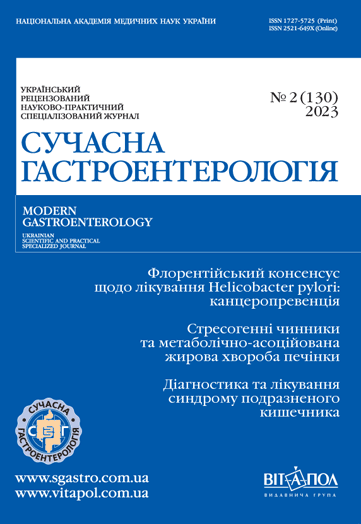
Вплив стресогенних чинників на перебіг метаболічно-асоційованої жирової хвороби печінки. Огляд
DOI:
https://doi.org/10.30978/MG-2023-2-69Ключові слова:
метаболічно‑асоційована жирова хвороба печінки, стрес, кишкова мікробіотаАнотація
Метаболічно‑асоційована жирова хвороба печінки (МАЖХП) є однією з найпоширеніших хвороб печінки у світі та зокрема в Україні. До комплексних патофізіологічних механізмів МАЖХП належать порушення різних процесів (інсулінорезистентність, ліпотоксичність, прозапальна активація, зміни кишкової мікробіоти тощо). Метаболічно‑асоційована жирова хвороба печінки тісно повязана з порушеннями ментального здоров’я, і вони потенціюють перебіг одне одного. Стрес індукує як поведінкові, так і біологічні реакції, які активують вісь гіпоталамус — гіпофіз — наднирники, що призводить до підвищення рівня кортизолу та прозапальних біомаркерів, які можуть бути залучені до розвитку МАЖХП. Даних щодо впливу стресогенних чинників воєнного та післявоєнного стану на формування і прогресуванння МАЖХП недостатньо. Мало відомо про роль біомаркерів хронічного стресу (дегідроепіандростерону, пролактину, нейротропного фактора головного мозку (BDNF)) у патогенезі МАЖХП. Суперечливі дані літератури щодо змін якісного та кількісного складу кишкової мікробіоти на рівні основних філів у хворих на МАЖХП. Не досліджено динаміки кишкової мікробіоти під впливом стресових чинників воєнного та післявоєнного часу. Комплексне визначення ступеня порушення цілісності епітеліального бар’єра кишечника, стану КМ, рівня гормонів стресу та прозапальних чинників у хворих на МАЖХП у стресогенних умовах воєнного і післявоєнного стану дасть змогу уточнити патофізіологічні ланки формування та прогресування МАЖХП і розробити патогенетично обґрунтовані профілактичні та терапевтичні втручання для гальмування розвитку запалення і процесів фіброзування в тканині печінки у хворих на МАЖХП шляхом корекції мікробіоти та модифікації стресових чинників воєнного та післявоєнного стану, а також дослідити вплив розробленої терапії на клінічні вияви, метаболічний профіль, склад КМ, запалення та рівень нейротропних гормонів у хворих на МАЖХП. Таким чином, наукові дослідження впливу стресогенних чинників на перебіг МАЖХП та визначення патофізіологічних механізмів є актуальними та доцільними.
Посилання
Fadieienko GD, Gridnyev OY, Chereliuk NI, Kurinna OG. The role of intestinal microbiome in the progression of non-alcoholic fatty liver disease. Modern Gastroenterology (Ukraine). 2019;(4):92-9. http://doi.org/10.30978/MG-2019-4-92.
Araújo J, Cai J, Stevens J. Prevalence of optimal metabolic health in American adults: National Health and Nutrition Examination Survey 2009-2016. Metabolic Syndrome and Related Disorders. 2019;17(1):46-52. http://doi.org/10.1089/met.2018.0105.
Branković M, Jovanović I, Dukić M, et al. Lipotoxicity as the leading cause of non-alcoholic steatohepatitis. International Journal of Molecular Sciences. 2022;23(9):5146. http://doi.org/10.3390/ijms23095146.
Chalasani N, Younossi Z, Lavine JE, et al. The diagnosis and management of nonalcoholic fatty liver disease: practice guidance from the American Association for the Study of Liver Diseases. Hepatology. 2018;67(1):328-57. http://doi.org/10.1002/hep.29367.
Chen Y-l, Li H, Li S, et al. Prevalence of and risk factors for metabolic associated fatty liver disease in an urban population in China: a cross-sectional comparative study. BMC Gastroenterology. 2021;21(1):212. http://doi.org/10.1186/s12876-021-01782-w.
Dutheil F, de Saint Vincent S, Pereira B, et al. DHEA as a biomarker of stress: a systematic review and meta-analysis. Frontiers in Psychiatry. 2021;12:1-14. http://doi.org/10.3389/fpsyt.2021.688367.
Eslam M, Newsome PN, Sarin SK, et al. A new definition for metabolic dysfunction-associated fatty liver disease: An international expert consensus statement. J Hepatol. 2020;73(1):202-9. http://doi.org/10.1016/j.jhep.2020.03.039.
Eslam M, Sanyal AJ, George J. MAFLD: A consensus-driven proposed nomenclature for metabolic associated fatty liver Disease. Gastroenterology. 2020;158(7):1999-2014.e1991. http://doi.org/10.1053/j.gastro.2019.11.312.
Fadieienko GD, Chereliuk NI, Galchinskaya VY. RATIO of main phylotypes of gut microbiota in patients with non-alcoholic fatty liver disease depending on the body mass index. Wiadomosci lekarskie (Warsaw, Poland). 2021;74 (3 cz 1):523-8.
Ferro D, Baratta F, Pastori D, et al. New insights into the pathogenesis of non-alcoholic fatty liver disease: gut-derived lipopolysaccharides and oxidative stress. Nutrients. 2020;12(9):2762. http://doi.org/10.3390/nu12092762.
Han AL. Association between Non-alcoholic fatty liver disease and dietary habits, stress, and health-related quality of life in Korean adults. Nutrients. 2020;12(6):1555. http://doi.org/10.3390/nu12061555.
Hattori Y, Yamada H, Munetsuna E, et al. Increased brain-derived neurotrophic factor in the serum of persons with nonalcoholic fatty liver disease. Endocrine Journal. 2022; 69 (8):999-1006. http://doi.org/10.1507/endocrj.EJ21-0584.
Inoue Y, Qin B, Poti J, Sokol R, Gordon-Larsen P. Epidemiology of obesity in adults: latest trends. Current Obesity Reports. 2018;7(4):276-88. http://doi.org/10.1007/s13679-018-0317-8.
Kang D, Zhao D, Ryu S, Guallar E, Cho J, Lazo M, Shin H, Chang Y, Sung E. Author Correction: Perceived stress and non-alcoholic fatty liver disease in apparently healthy men and women. Sci Rep. 2020 Dec 9;10(1):21978. http://doi.org/10.1038/s41598-020-78911-0. Erratum for: Sci Rep. 2020 Jan 8;10(1):38.
Kessoku T, Kobayashi T, Tanaka K, et al. The role of leaky gut in nonalcoholic fatty liver disease: a novel therapeutic target. International Journal of Molecular Sciences. 2021;22 (15):8161. http://doi.org/10.3390/ijms22158161.
Koga M, Saito H, Mukai M, Saibara T, Kasayama S. Serum dehydroepiandrosterone sulphate levels in patients with non-alcoholic fatty liver disease. Internal Medicine (Tokyo, Japan). 2011;50 (16):1657-61. http://doi.org/10.2169/internalmedicine.50.4682.
Lau LHS, Wong SH. Microbiota, Obesity and NAFLD. Advances in Experimental Medicine and Biology. 2018;1061:111-25. http://doi.org/10.1007/978-981-10-8684-7_9.
Liu W, Han X, Zhou X, et al. Brain derived neurotrophic factor in newly diagnosed diabetes and prediabetes. Molecular and Cellular Endocrinology. 2016;429:106-13. http://doi.org/10.1016/j.mce.2016.04.002.
Mason BN, Kallianpur R, Price TJ, Akopian AN, Dussor GO. Prolactin signaling modulates stress-induced behavioral responses in a preclinical mouse model of migraine. Headache. 2022;62(1):11-25. http://doi.org/10.1111/head.14248.
Mikolasevic I, Delija B, Mijic A, et al. Small intestinal bacterial overgrowth and non-alcoholic fatty liver disease diagnosed by transient elastography and liver biopsy. International Journal of Clinical Practice. 2021;75(4):e13947. http://doi.org/10.1111/ijcp.13947.
Milić S, Lulić D, Štimac D. Non-alcoholic fatty liver disease and obesity: biochemical, metabolic and clinical presentations. World Journal of Gastroenterology. 2014;20 (28):9330-7.
Monteleone P, Tortorella A, Martiadis V, Serritella C, Fuschino A, Maj M. Opposite changes in the serum brain-derived neurotrophic factor in anorexia nervosa and obesity. Psychosomatic Medicine. 2004;66(5):744-8. http://doi.org/10.1097/01.psy.0000138119.12956.99.
Mouzaki M, Comelli EM, Arendt BM, Bonengel J, Fung SK, Fischer SE, McGilvray ID, Allard JP. Intestinal microbiota in patients with nonalcoholic fatty liver disease. Hepatology. 2013 Jul;58(1):120-7. http://doi.org/10.1002/hep.26319. Epub 2013 May 14. PMID: 23401313.
Murakami S, Imbe H, Morikawa Y, Kubo C, Senba E. Chronic stress, as well as acute stress, reduces BDNF mRNA expression in the rat hippocampus but less robustly. Neuroscience Research. 2005;53(2):129-39. http://doi.org/10.1016/j.neures.2005.06.008.
Nguyen VH, Le MH, Cheung RC, Nguyen MH. Differential clinical characteristics and mortality outcomes in persons with NAFLD and/or MAFLD. Clinical Gastroenterology and Hepatology. 2021;19 (10):2172-81.e2176. http://doi.org/10.1016/j.cgh.2021.05.029.
Russ TC, Kivimäki M, Morling JR, Starr JM, Stamatakis E, Batty GD. Association between psychological distress and liver disease mortality: a meta-analysis of individual study participants. Gastroenterology. 2015;148(5):958-66.e954. http://doi.org/10.1136/bmj.e4933.
Sakurai Y, Kubota N, Yamauchi T, Kadowaki T. Role of insulin resistance in MAFLD. International Journal of Molecular Sciences. 2021;22(8):4156. http://doi.org/10.3390/ijms22084156.
Scarpellini E, Abenavoli L, Cassano V, Rinninella E, Sorge M, Capretti F, Rasetti C, Svegliati Baroni G, Luzza F, Santori P, Sciacqua A. The Apparent Asymmetrical Relationship Between Small Bowel Bacterial Overgrowth, Endotoxemia, and Liver Steatosis and Fibrosis in Cirrhotic and Non-Cirrhotic Patients: A Single-Center Pilot Study. Front Med (Lausanne). 2022 Apr 26;9:872428. http://doi.org/10.3389/fmed.2022.872428. PMID: 35559337; PMCID: PMC9090439.
Schwenger KJP, Fischer SE, Jackson TD, Okrainec A, Allard JP. Non-alcoholic fatty liver disease in morbidly obese individuals undergoing bariatric surgery: prevalence and effect of the pre-bariatric very low calorie diet. Obesity Surgery. 2018;28(4):1109-16. http://doi.org/10.1007/s11695-017-2980-3.
Shao S, Yao Z, Lu J, et al. Ablation of prolactin receptor increases hepatic triglyceride accumulation. Biochemical and Biophysical Research Communications. 2018;498(3):693-9. http://doi.org/10.1016/j.bbrc.2018.03.048.
Shea S, Lionis C, Kite C, et al. Non-alcoholic fatty liver disease (NAFLD) and Potential links to depression, anxiety, and chronic stress. Biomedicines. 2021;9 (11):1697. http://doi.org/10.3390/biomedicines9111697.
Sobhonslidsuk A, Chanprasertyothin S, Pongrujikorn T, Kaewduang P, Promson K, Petraksa S, Ongphiphadhanakul B. The Association of Gut Microbiota with Nonalcoholic Steatohepatitis in Thais. Biomed Res Int. 2018 Jan 16;2018:9340316. http://doi.org/10.1155/2018/9340316. PMID: 29682571; PMCID: PMC5842744.
Stefan N, Schick F, Häring HU. Causes, characteristics, and consequences of metabolically unhealthy normal weight in humans. Cell Metabolism. 2017;26(2):292-300. http://doi.org/10.1016/j.cmet.2017.07.008.
Tilg H, Effenberger M. From NAFLD to MAFLD: when pathophysiology succeeds. Nat Rev Gastroenterol Hepatol. 2020 Jul;17(7):387-388. http://doi.org/10.1038/s41575-020-0316-6. Epub 2020 May 27. PMID: 32461575.
Wong VW, Tse CH, Lam TT, et al. Molecular characterization of the fecal microbiota in patients with nonalcoholic steatohepatitis-a longitudinal study. PloS One. 2013;8(4):e62885. http://doi.org/10.1371/journal.pone.0062885.
Woodhouse CA, Patel VC, Singanayagam A, Shawcross DL. Review article: the gut microbiome as a therapeutic target in the pathogenesis and treatment of chronic liver disease. Alimentary Pharmacology & Therapeutics. 2018;47(2):192-202. http://doi.org/10.1111/apt.14397.
Younossi Z, Anstee QM, Marietti M, et al. Global burden of NAFLD and NASH: trends, predictions, risk factors and prevention. Nature reviews Gastroenterology & Hepatology. 2018;15(1):11-20. http://doi.org/10.1038/nrgastro.2017.109.
Zhang P, Feng W, Chu X, Sun X, Zhu D, Bi Y. A newly noninvasive model for prediction of non-alcoholic fatty liver disease: utility of serum prolactin levels. BMC Gastroenterology. 2019;19(1):202. http://doi.org/10.1186/s12876-019-1120-z
##submission.downloads##
Опубліковано
Номер
Розділ
Ліцензія
Авторське право (c) 2023 Автор

Ця робота ліцензується відповідно до Creative Commons Attribution-NoDerivatives 4.0 International License.


