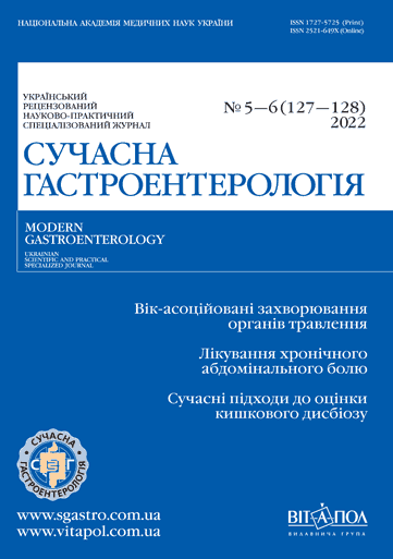
Сучасні підходи до суті та оцінки кишкового дисбіозу. Огляд
DOI:
https://doi.org/10.30978/MG-2022-5-58Ключові слова:
кишковий дисбіоз, індекси дисбіозуАнотація
Дисбаланс, дисфункція або порушення кишкової мікробіоти дедалі частіше визнають як індикатор певного захворювання або поганого стану здоров’я. Через складність та індивідуальні варіації в мікробних угрупованнях відсутній золотий стандарт для визначення наявності або ступеня цього дисбалансу чи порушення кишкової мікробіоти, хоча в багатьох дослідженнях це позначають терміном «кишковий дисбіоз». Проблема з визначенням кишкового дисбіозу зумовлена відсутністю чіткого визначення здорової кишкової мікробіоти. Для кваліфікації терміну «кишковий дисбіоз» визначено та застосовано кілька індексів. Згідно з результатами пошуку великого масиву літератури, відомі індекси дисбіозу методологічно можна згрупувати в п’ять категорій: масштабне профілювання бактеріальних маркерів, релевантні методи на основі таксонів, класифікація оточення, випадковий прогноз, комбінована α/β‑різноманітність. Наведені методи визначення та кількісної оцінки кишкового дисбіозу успішно фіксують відмінності між кишковою мікробіотою, пов’язаною зі специфічними станами, захворюваннями або втручаннями, а також наявною у здорових осіб або на початку лікування (до втручання). Такі індекси можуть допомогти схарактеризувати захворювання та несприятливі стани, спрогнозувати результати лікування та надати інформацію, відмінну від загальноприйнятих α‑ та β‑оцінок мікробної різноманітності. Слід пам’ятати, що дисбіоз не є чітко визначеним станом і що індекси дисбіозу відрізняються за методологічними та клінічними контекстами, розроблені для різних когорт і опису різноманітних станів. Висвітлено сучасні основні методи та принципи, пов’язані з оцінкою кишкової мікробіоти у клінічному контексті, зокрема застосування індексів дисбіозу, їхній розрахунок та показники в контексті специфічних захворювань і станів. Індекси дисбіозу не є окремим вимірюванням, їх слід інтерпретувати в контексті клінічних даних. Слід пам’ятати, що кишкова мікробіота, що характеризує певне захворювання або втручання, часто змінюється внаслідок порушення таких чинників, як дієта, прийом лікарських препаратів, доступність кисню або імунні реакції. У такому випадку індекси дисбіозу застосовують як діагностичний маркер, але не обов’язково як предиктор захворювання. Значення індексів дисбіозу як простих інструментів для опису відмінностей між різними кишковими мікробними угрупованнями є важливим.
Посилання
Aasbrenn M., Valeur J., Farup P. G. Evaluation of a faecal dysbiosis test for irritable bowel syndrome in subjects with and without obesity. Scand J Clin Lab Invest. 2018;78:109-113. https://doi.org/10.1080/00365513.2017.1419372.
Bajaj J. S., Heuman D. M., Hylemon P. B. et al. Altered profile of human gut microbiome is associated with cirrhosis and its complications. J Hepatol. 2014;60:940-947. https://doi.org/10.1016/j.jhep.2013.12.019.
Bennet S. M. P., Böhn L., Störsrud S. et al. Multivariate modelling of faecal bacterial profiles of patients with IBS predicts responsiveness to a diet low in FODMAPs. Gut. 2018;67:872-881. https://doi.org/10.1136/gutjnl-2016-313128.
Brahe L. K., Le Chatelier E., Prifti E. et al. Dietary modulation of the gut microbiota–a randomised controlled trial in obese postmenopausal women. Br J Nutr. 2015;114:406-417. https://doi.org/10.1017/S0007114515001786.
Casén C., Vebø H. C., Sekelja M. et al. Deviations in human gut microbiota: a novel diagnostic test for determining dysbiosis in patients with IBS or IBD. Aliment Pharmacol Ther. 2015;42:71-83. https://doi.org/10.1111/apt.13236.
Castaner O., Goday A., Park Y. M. et al. The gut microbiome profile in obesity: a systematic review. Int J Endocrinol. 2018;2018:1-9. https://doi.org/10.1155/2018/4095789.
Chen L., Reeve J., Zhang L., Huang S., Wang X., Chen J. GMPR: a robust normalization method for zero-inflated count data with application to microbiome sequencing data. Peer J 2018. 6. e4600. https://doi.org/10.7717/peerj.4600./.
El-Salhy M., Hausken T., Hatlebakk J. G. Increasing the dose and/or repeating faecal microbiota transplantation (FMT) increases the response in patients with irritable bowel syndrome (IBS). Nutrients. 2019;11. 1415. https://doi.org/10.3390/nu11061415.
Falony G., Joossens M., Vieira-Silva S. et al. Population-level analysis of gut microbiome variation. Science. 2016;352:560-564. https://doi.org/10.1126/science.aad3503.
Farup P. G., Aasbrenn M., Valeur J. Separating «good» from «bad» faecal dysbiosis — evidence from two cross-sectional studies. BMC Obes. 2018;5. 30. https://doi.org/10.1186/s40608-018-0207-3.
Farup P. G., Lydersen S., Valeur J. Are nonnutritive sweeteners obesogenic? Associations between diet, faecal microbiota, and short-chain fatty acids in morbidly obese subjects. J Obes. 2019;4608315-26. https://doi.org/10.1155/2019/4608315.
Farup P. G., Valeur J. Changes in faecal short-chain fatty acids after weight-loss interventions in subjects with morbid obesity. Nutrients. 2020;12:802-814. https://doi.org/10.3390/nu12030802.
Ferrannini E. The target of metformin in type 2 diabetes. N Engl J Med. 2014;371:1547-1548. https://doi.org/10.1056/NEJMcibr1409796.
Forslund K., Hildebrand F., Nielsen T. et al., MetaHIT Consortium. Disentangling type 2 diabetes and metformin treatment signatures in the human gut microbiota. Nature. 2015;528:262-266. https://doi.org/10.1038/nature15766.
Franzosa E. A., McIver L. J., Rahnavard G. et al. Species-level functional profiling of metagenomes and metatranscriptomes. Nat Methods. 2018;15:962-968. https://doi.org/10.1038/s41592-018-0176-y.
Gevers D., Kugathasan S., Denson L. A. et al. The treatment-naive microbiome in new-onset Crohn’s disease. Cell Host Microbe. 2014;15:382-392. https://doi.org/10.1016/j.chom.2014.02.005.
Huang S., Li R., Zeng X. et al. Predictive modeling of gingivitis severity and susceptibility via oral microbiota. ISME J. 2014;8:1768-1780. https://doi.org/10.1038/ismej.2014.32.
Hustoft T. N., Hausken T., Ystad S. O. et al. Effects of varying dietary content of fermentable short-chain carbohydrates on symptoms, fecal microenvironment, and cytokine profiles in patients with irritable bowel syndrome. Neurogastroenterol Motil. 2017;29. e12969. https://doi.org/10.1111/nmo.12969.
Jeffery I. B., O’Toole P.W., Öhman L. et al. An irritable bowel syndrome subtype defined by species specific alterations in faecal microbiota. Gut. 2012;61:997-1006. https://doi.org/10.1136/gutjnl-2011-301501.
Jørgensen S. F., Trøseid M., Kummen M. et al. Altered gut microbiota profile in common variable immunodeficiency associates with levels of lipopolysaccharide and markers of systemic immune activation. Mucosal Immunol. 2016;9:1455-1465. https://doi.org/10.1038/mi.2016.18.
Khor B., Gardet A., Xavier R. J. Genetics and pathogenesis of inflammatory bowel disease. Nature. 2011;474:307-317. https://doi.org/10.1038/nature10209.
Kim Y. J., Choi Y. S., Baek K. J. et al. Mucosal and salivary microbiota associated with recurrent aphthous stomatitis. BMC Microbiol. 2016;16:1-10. https://doi.org/10.1186/s12866-016-0673-z.
Le Chatelier E., Nielsen T., Qin J. et al., MetaHIT Consortium. Richness of human gut microbiome correlates with metabolic markers. Nature. 2013;500:541-546. https://doi.org/10.1038/nature12506.
Levy M., Kolodziejczyk A. A., Thaiss C. A., Elinav E. Dysbiosis and the immune system. Nat Rev Immunol. 2017;17:219-232. https://doi.org/10.1038/nri.2017.7.
Ley R. E., Turnbaugh P. J., Klein S., Gordon J. I. Microbial ecology: human gut microbes associated with obesity. Nature. 2006;444:1022-1023. https://doi.org/10.1038/4441022a.
Liu Y., Jin Y., Li J. et al. Small bowel transit and altered gut microbiota in patients with liver cirrhosis. Front Physiol. 2018;9. 470. https://doi.org/10.3389/fphys.2018.00470.
Lloyd-Price J., Arze C., Ananthakrishnan A. N. et al., IBD MDB Investigators. Multi-omics of the gut microbial ecosystem in inflammatory bowel diseases. Nature. 2019;569:655-662. https://doi.org/10.1038/s41586-019-1237-9.
Magnusson M. K., Strid H., Sapnara M. et al. Anti-TNF therapy response in patients with ulcerative colitis is associated with colonic antimicrobial peptide expression and microbiota composition. J Crohns Colitis. 2016;10:943-952. https://doi.org/10.1093/ecco-jcc/jjw051.
Mandl T., Marsal J., Olsson P., Ohlsson B., Andréasson K. Severe intestinal dysbiosis is prevalent in primary Sjögren’s syndrome and is associated with systemic disease activity. Arthritis Res Ther. 2017;19. 237. https://doi.org/10.1186/s13075-017-1446-2.
Manichanh C., Borruel N., Casellas F., Guarner F. The gut microbiota in IBD. Nat Rev Gastroenterol Hepatol. 2012;9:599-608. https://doi.org/10.1038/nrgastro.2012.152.
Mayerhofer C. C. K., Kummen M., Holm K. et al. Low fibre intake is associated with gut microbiota alterations in chronic heart failure. ESC Hear Fail. 2020;7:456-466. https://doi.org/10.1002/ehf2.12596.
Mazzawi T., Lied G. A., Sangnes D. A. et al. The kinetics of gut microbial community composition in patients with irritable bowel syndrome following fecal microbiota transplantation. PLoS One. 2018;13. e0194904-17. https://doi.org/10.1371/journal.pone.0194904.
Montassier E., Al-Ghalith G. A., Hillmann B. et al. CLOUD: a non-parametric detection test for microbiome outliers. Microbiome. 2018;6. 137. https://doi.org/10.1186/s40168-018-0514-4.
Nakatsu G., Li X., Zhou H. et al. Gut mucosal microbiome across stages of colorectal carcinogenesis. Nat Commun. 2015;6:30-32. https://doi.org/10.1038/ncomms9727.
Olesen S. W., Alm E. J. Dysbiosis is not an answer. Nat Microbiol. 2016;1. 16228. https://doi.org/10.1038/nmicrobiol.2016.228.
Saffouri G. B., Shields-Cutler R.R., Chen J. et al. Small intestinal microbial dysbiosis underlies symptoms associated with functional gastrointestinal disorders. Nat Commun. 2019;10. 2012. https://doi.org/10.1038/s41467-019-09964-7.
Santiago M., Eysenbach L., Allegretti J. et al. Microbiome predictors of dysbiosis and VRE decolonization in patients with recurrent C. difficile infections in a multi-center retrospective study. AIMS Microbiol. 2019;5:1-18. https://doi.org/10.3934/microbiol.2019.1.1.
Sarangi A. N., Goel A., Aggarwal R. Methods for studying gut microbiota: a primer for physicians. J Clin Exp Hepatol. 2019;9:62-73. https://doi.org/10.1016/j.jceh.2018.04.016.
Shen Y., Xu J., Li Z. et al. Analysis of gut microbiota diversity and auxiliary diagnosis as a biomarker in patients with schizophrenia: a cross-sectional study. Schizophr Res. 2018;197:470-477. https://doi.org/10.1016/j.schres.2018.01.002.
Soares R. C., Camargo-Penna P.H., De Moraes V. C. S. et al. Dysbiotic bacterial and fungal communities not restricted to clinically affected skin sites in dandruff. Front Cell Infect Microbiol. 2016;6. 157. https://doi.org/10.3389/fcimb.2016.00157.
Sokol H., Leducq V., Aschard H. et al. Fungal microbiota dysbiosis in IBD. Gut. 2017;66:1039-1048. https://doi.org/10.1136/gutjnl-2015-310746.
Sze M. A., Schloss P. D. Looking for a signal in the noise: revisiting obesity and the microbiome. mBio. 2016;7:e01018-16. https://doi.org/10.1128/mBio.01018-16.
Thorsen J., Brejnrod A., Mortensen M. et al. Large-scale benchmarking reveals false discoveries and count transformation sensitivity in 16S rRNA gene amplicon data analysis methods used in microbiome studies. Microbiome. 2016;4. 62. https://doi.org/10.1186/s40168-016-0208-8.
Truong D. T., Franzosa E. A., Tickle T. L. et al. MetaPhlAn2 for enhanced metagenomic taxonomic profiling. Nat Methods. 2015;12:902-903. https://doi.org/10.1038/nmeth.3589.
Valeur J., Småstuen M. C., Knudsen T., Lied G. A., Røseth A. G. Exploring gut microbiota composition as an indicator of clinical response to dietary FODMAP restriction in patients with irritable bowel syndrome. Dig Dis Sci. 2018;63:429-436. https://doi.org/10.1007/s10620-017-4893-3.
Vich Vila A., Collij V., Sanna S. et al. Impact of commonly used drugs on the composition and metabolic function of the gut microbiota. Nat Commun. 2020;11:1-11. https://doi.org/10.1038/s41467-019-14177-z.
Wang J., Wang Y., Zhang X. et al. Gut microbial dysbiosis is associated with altered hepatic functions and serum metabolites in chronic hepatitis B patients. Front Microbiol. 2017;8. 2222. https://doi.org/10.3389/fmicb.2017.02222.
Wei S., Bahl M. I., Baunwall S. M. D., Hvas C. L., Licht T. R. Determining gut microbial dysbiosis: a review of applied indexes for assessment of intestinal microbiota imbalances. Appl Environ Microbiol. 2021;87. e00395-21. https://doi.org/10.1128/AEM.00395-21.
##submission.downloads##
Опубліковано
Номер
Розділ
Ліцензія
Авторське право (c) 2022 Автори

Ця робота ліцензується відповідно до Creative Commons Attribution-NoDerivatives 4.0 International License.


