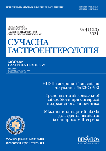
Сучасні уявлення про стан епітеліального бар’єра стравоходу при гастроезофагеальній рефлюксній хворобі та можливості корекції за допомогою альгінатів
DOI:
https://doi.org/10.30978/MG-2021-4-13Ключові слова:
гастроезофагеальна рефлюксна хвороба, проникність епітелію, бар’єрна функція, щільні контакти, альгінатиАнотація
Гастроезофагеальна рефлюксна хвороба (ГЕРХ) залишається актуальною проблемою клінічної медицини. Пошук шляхів ефективного лікування зумовлює необхідність розставлення нових акцентів у мультифакторному патогенезі захворювання. Відповідно до сучасних тенденцій особливу увагу приділяють епітеліальному бар’єру стравоходу. В статті розглянуто структуру епітеліального шару слизової оболонки, особливості транспортних шляхів і міжклітинних взаємодій епітелію стравоходу. Особливу увагу приділено феномену розширення міжклітинних просторів (РМП) і синдрому підвищеної епітеліальної проникності (СПЕП) при ГЕРХ. Дано трактування цих понять, розкрито їхню роль у реалізації запальних процесів і клінічних виявів захворювання, таких як печія та біль. Наведено дані про можливі механізми формування феномену РМП і СПЕП, методи їхньої діагностики та аспекти, які обговорюються в науковому світі.
Показано можливість цитопротекторної терапії за допомогою застосування альгінатних препаратів. На фармацевтичному ринку України з альгінатів представлено фармакологічну лінійку Гавіскон® («Реккітт Бенкізер Україна», Велика Британія). Наведено результати експериментальних досліджень з використанням новітніх технологій і дані рандомізованих клінічних досліджень, що підтверджують клінічну ефективність альгінатів.
Феномен РМП епітелію стравоходу та СПЕП для чинників агресії — одна з провідних ланок патогенезу ГЕРХ, зокрема її неерозивної форми, та певною мірою визначає особливості клінічних виявів захворювання. Лікування ГЕРХ із застосуванням альгінатів обґрунтоване, його успішно використовують у клінічній практиці та активно вивчають. Використання препаратів фармакологічної лінійки Гавіскон® є доцільним завдяки їхньому унікальному механізмові дії із забезпеченням підвищення резистентності слизової оболонки стравоходу.
Посилання
Shcherbуnina MB, Solovіovа NE. A strategy to protect the integrity of the esophageal mucosa in the treatment of gastroesophageal reflux disease [in Urkainian]. Modern Gastroenterology. 2020;1:60-. http://doi.org/10.30978/MG-2020-1-60.
Altomare A, Luca Guarino Sara Emerenziani MP, Cicala M et al. Gastrointestinal sensitivity and gastroesophageal reflux disease. Ann N Y Acad Sci. 2013;1300:80-95. doi: 10.1111/nyas.12236.
Azumi T, Adachi K, Furuta K et al. Esophageal epithelial surface in patients with gastroesophageal reflux disease: an electron microscopic study. World J Gastroenterol. 2008;14 (37):5712-5716. doi: 10.3748/wjg.14.5712.
Björkman EV, Edebo A, Oltean M, Casselbrant A. Esophageal barrier function and tight junction expression in healthy subjects and patients with gastroesophageal reflux disease: functionality of esophageal mucosa exposed to bile salt and trypsin in vitro. Scand J Gastroenterol. 2013;48 (10):1118-1126. doi: 10.3109/00365521.2013.828772.
Blevins C, Dierkhising D, Geno D, Johnson M. Obesity and GERD impair esophageal epithelial permeability through 2 distinct mechanisms. Neurogastroenterol Motil. 2018;30 (10):e13403. doi: 10.1111/nmo.13403.
Caviglia R, Ribolsi M, Gentile M et al. Dilated intercellular spaces and acid reflux at the distal and proximal oesophagus in patients with non-erosive gastro-oesophageal reflux disease. Aliment Pharmacol Ther. 2007;25(5):629-636. doi: 10.1111/j.1365-2036.2006.03237.
Dent J. Patterns of lower esophageal sphincter function associated with gastroesophageal reflux. Am J Med. 1997;103 (5A):29S-32S. doi: 10.1016/s0002-9343 (97)00317-3.
Deraman MA, Abdul Hafidz MI, Lawenko RM et al. Randomised clinical trial: the effectiveness of Gaviscon Advance vs non-alginate antacid in suppression of acid pocket and post-prandial reflux in obese individuals after late-night supper. Aliment Pharmacol Ther. 2020;51 (11):1014-1021. doi: 10.1111/apt.15746. Epub 2020 Apr 28. PMID: 32343001. PMCID: PMC7318318.
Farré R, De Vos R, Geboes K et al. Critical role of stress in increased oesophageal mucosa permeability and dilated intercellular spaces. Gut. 2007;56(9):1191-1197. doi: 10.1136/gut.2006.113688.
France MM, Turner JR. The mucosal barrier at a glance. J Cell Sci. 2017;130(2):307-314. doi: 10.1242/jcs.193482.
Gyawali CP, Kahrilas PJ, Savarino E et al. Modern diagnosis of GERD: the Lyon Consensus. Gut. 2018;67(7):1351-1362. doi: 10.1136/gutjnl-2017-314722.
Herregods TV, Bredenoord AJ, Smout AJ. Pathophysiology of gastroesophageal reflux disease: new understanding in a new era. Neurogastroenterol Motil. 2015;27(9):1202-1213. doi: 10.1111/nmo.12611.
Ito H, Iijima K, Ara N et al. Reactive nitrogen oxide species induce dilatation of the intercellular space of rat esophagus. Scand J Gastroenterol. 2010;45(3):282-291. doi: 10.3109/00365520903469956.
Kahrilas PJ. Dilated intercellular spaces: extending the reach of the endoscope. Am J Gastroenterol. 2005;100(3):549-550. doi: 10.1111/j.1572-0241.2005.41918.x.
Malenstein H, Farré R, Sifrim D. Esophageal dilated intercellular spaces (DIS) and nonerosive reflux disease. Am J Gastroenterol. 2008;103(4):1021-1028. doi: 10.1111/j.1572-0241.2007.01688.x.
Mönkemüller K, Wex T, Kuester D et al. Role of tight junction proteins in gastroesophageal reflux disease. BMC Gastroenterol. 2012;12:128. doi: 10.1186/1471-230X-12-128.
Odenwald MA, Turner JR. The intestinal epithelial barrier: a therapeutic target?. Nat RevGastroenterol Hepatol. 2017;14(1):9-21. doi: 10.1038/nrgastro.2016.169.
Orlando LA, Orlando RC. Dilated intercellular spaces as a marker of GERD. Curr Gastroenterol Rep. 2009;11(3):190-194. doi: 10.1007/s11894-009-0030-6. PMID: 19463218.
Orlando RC. Dilated intercellular spaces and chronic cough as an extra-oesophageal manifestation of gastrooesophageal reflux disease. Pulm Pharmacol Ther. 2011;24(3):272-275. doi: 10.1016/j.pupt.2010.10.007.
Souza RF, Huo X, Mittal V et al. Gastroesophageal reflux might cause esophagitis through a cytokine-mediated mechanism rather than caustic acid injury. Gastroenterology. 2009;137(5):1776-1784. doi: 10.1053/j.gastro.2009.07.055.
Strugala V, Avis J, Jolliffe IG et al. The role of an alginate suspension on pepsin and bile acids — key aggressors in the gastric refluxate. Does this have implications for the treatment of gastro-oesophageal reflux disease?. J Pharm Pharmacol. 2009;61:1021-1028.
Suzuki H, Iijima K, Scobie G, Fyfe V, McColl KE. Nitrate and nitrosative chemistry within Barrett’s oesophagus during acid reflux. Gut. 2005;54 (11):1527-3155. doi: 10.1136/gut.2005.066043.
Tobey NA, Carson JL, Alkiek RA, Orlando RC. Dilated intercellular spaces: a morphological feature of acid reflux — damaged human esophageal epithelium. Gastroenterology. 1996;111(5):1200-1205. doi: 10.1053/gast.1996.v111.pm8898633.
Tobey NA, Gambling TM, Vanegas XC, Carson JL.., Orlando R. C. Physicochemical basis for dilated intercellular spaces in non-erosive acid-damaged rabbit esophageal epithelium. Dis Esophagus. 2008;21(8):757-764. doi: 10.1111/j.1442-2050.2008.00841.x.
Weber CR, Raleigh DR, Su L et al. Epithelial myosin light chain kinase activation induces mucosal interleukin-13 expression to alter tight junction ion selectivity. J Biol Chem. 2010;285 (16):12037-12046. doi: 10.1074/jbc.M109.064808.
Weijenborg PW, Smout AJ, Verseijden C et al. Hypersensitivity to acid is associated with impaired esophageal mucosal integrity in patients with gastroesophageal reflux disease with and without esophagitis. Am J Physiol. Gastrointest. Liver Physiol. 2014;307(3):G323-329. doi: 10.1152/ajpgi.00345.2013.
Wilkinson J, Wade A, Thomas SJ et al. Randomized clinical trial: a double-blind, placebo-controlled study to assess the clinical efficacy and safety of alginate-antacid (Gaviscon Double Action) chewable tablets in patients with gastro-oesophageal reflux disease. Eur J Gastroenterol Hepatol. 2019;31(1):86-93. doi: 10.1097/MEG.0000000000001258. PMID: 30272584.
Woodland P, Lee C, Duraisamy Y et al. Assessment and protection of esophageal mucosal integrity in patients with heartburn without esophagitis. Am J Gastroenterol. 2013;108:535-543. doi: 10.1038/ajg.2013.101.


