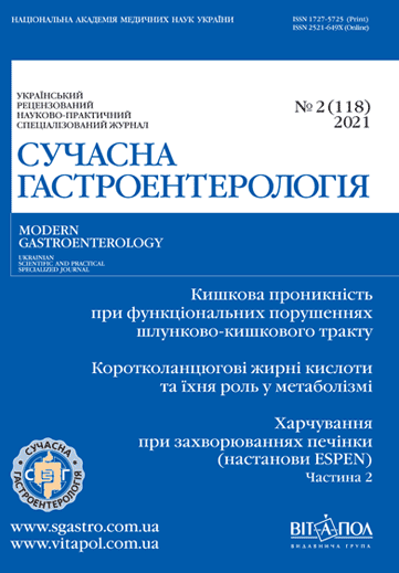
Етіопатогенетичні аспекти формування функціональних захворювань шлунково-кишкового тракту: акцент на зміни кишкової проникності
DOI:
https://doi.org/10.30978/MG-2021-2-79Ключові слова:
синдром подразненого кишечника, функціональна диспепсія, кишечна проникність, цитокіни, трансмембранні білкиАнотація
Синдром подразненого кишечника (СПК) та функціональна диспепсія (ФД) належать до найпоширеніших патологій органів травлення. Проаналізовано дані літератури щодо порушення кишкової проникності як етіопатогенетичного чинника розвитку функціональних захворювань шлунково‑кишкового тракту (ШКТ).
Слизова оболонка кишечника становить собою бар’єр, який виконує багато функцій, насамперед — захисну, а саме запобігає переходу внутрішньо просвітних субстанцій у внутрішнє середовище організму. Останнім часом накопичується дедалі більше наукових доказів того, що порушення проникності слизової оболонки кишечника є важливим чинником розвитку і прогресування функціональних захворювань ШКТ, зокрема СПК і ФД. При цих патологіях відзначено послаблення бар’єрної функції слизової оболонки кишечника внаслідок змін вмісту білків щільних контактів клітин, що підвищує проникність, стимулює перехід патогенних чинників з просвіту кишечника у власну пластинку слизової оболонки кишечника та спричиняє активацію імунокомпетентних клітин. У патогенезі СПК провідну роль відводять активації опасистих клітин у слизовій оболонці як тонкої, так і товстої кишки, а при ФД — опасистим клітинам та еозинофілам слизової оболонки дванадцятипалої кишки. Опасисті клітини, які належать до імунної системи, у відповідь на активацію виробляють запальні цитокіни, дія яких спрямована на зміну чутливості нервових закінчень, розташованих у слизовій оболонці, що спричиняє феномен вісцеральної гіперчутливості та зміну тонуса і моторної функції ШКТ.
Корекція змін кишкової проникності слизової оболонки ШКТ — потенційно одне з напрямів у лікуванні СПК і ФД. Необхідно провести дослідження ефективності препаратів, які впливають на функціонування кишкової стінки, щодо нормалізації кишкової проникності при захворюваннях ШКТ.
Посилання
Andreev DN, Zaborovskii AV, Trukhmanov AS et al. Evoliutsiia predstavlenii o funktsional’nykh zabolevaniiakh zheludochno-kishechnogo trakta v svete Rimskikh kriteriev IV peresmotra (2016 g.). RZhGGK. 2017;1:4-11 [in Russian].
Vialov SS. Mucosal permeability disturbances as a pathogenesis factor of gastrointestinal tract functional disorders: rationale and correction possibilities. Consilium Medicum. 2018;20 (12):99-104. DOI:10.26442/20751753.2018.12.180062 [in Russian].
Grigor’ev P. Ja., Jakovenko JeP. Metody korrekcii narushenij normal’nogo sostava kishechnogo biocenoza 13-11-2011. URL:http://www.eurolab-portal.ru/encyclopedia/565/46097/) [in Russian].
Ugolev AM. i dr. Vzaimootnoshenija fermentativnyh funkcij podzheludochnoj zhelezy i tonkoj kishki pri adaptivnyh processah. Fiziol Zhurn SSSR. 1978;T. 64(9):1221-1228 [in Russian].
Ugolev AM. i dr. Pishhevaritel’nye fermenty v zheludochno-kishechnom trakte, pochke, pecheni, selezenke pri razlichnyh funkcional’nyh sostojanijah. Ros Fiziol Zhurn. 1992;T. 78(9):76-83 [in Russian].
Ugolev AM. Jevoljucija pishhevarenija i principy jevoljucii funkcij. Jelementy sovremennogo funkcionalizma. L.: Nauka; 1985. 544 p [in Russian].
Ugolev A.M, Iezuitova HH, Timofeeva NM. Jenzimaticheskij bar’er tonkoj kishki. Fiziol Zhurn im I M Sechenova. 1992;T. 78(8):1-20 [in Russian].
Adamsson J, Ottsjö LS, Lundin SB et al. Gastric expression of IL-17A and IFNγ in Helicobacter pylori infected individuals is related to symptoms. Cytokine. 2017;99:30-34.
Bashashati M, Moossavi S, Cremon C et al. Colonic immune cells in irritable bowel syndrome: A systematic review and meta-analysis. Neurogastroenterol Motil. 2018;30(1). 3192.
Bashashati M, Moradi M, Sarosiek I. Interleukin-6 in irritable bowel syndrome: A systematic review and meta-analysis of IL-6 (-G174C) and circulating IL-6 levels. Cytokine. 2017;99. P. 132-138.
Bennet SM, Polster A, Törnblom H et al. Global cytokine profiles and association with clinical characteristics in patients with irritable bowel syndrome. Am J Gastroenterol. 2016;111(8):1165-1176.
Bertiaux-Vandaele N, Youmba SB, Belmonte L et al. The expression and the cellular distribution of the tight junction proteins are altered in irritable bowel syndrome patients with differences according to the disease subtype. Am J Gastroenterol. 2011;106 (12):2165-2173. doi: 10.1038/ajg.2011.257.
Binesh F, Akhondei M, Pourmirafzali H, Rajabzadeh Y. Determination of relative frequency of eosinophils and mast cells in gastric and duodenal mucosal biopsies in adults with non-ulcer dyspepsia. J Coll Physicians Surg Pak. 2013;23(5):326-329.
Bischoff SC, Barbara G, Buurman W et al. Intestinal permeability — a new target for disease prevention and therapy. BMC Gastroenterol. 2014;14:189.
Boyer J, Saint-Paul MC, Dadone B et al. Inflammatory cell distribution in colon mucosa as a new tool for diagnosis of irritable bowel syndrome: A promising pilot study. Neurogastroenterol Motil. 2018;30(1). 13223.
Camara-Lemarroy CR, Metz L, Meddings JB et al. The intestinal barrier in multiple sclerosis: implications for pathophysiology and therapeutics. Brain. 2018;141(7):1900-1916.
Choghakhori R, Abbasnezhad A, Hasanvand A, Amani R. Inflammatory cytokines and oxidative stress biomarkers in irritable bowel syndrome: Association with digestive symptoms and quality of life. Cytokine. 2017;93:34-43.
Choung RS. Natural history and overlap of functional gastrointestinal disorders. Korean J Gastroenterol. 2012;60(6):345-348.
Coeffier M, Gloro R, Boukhettala N, Aziz M, Lecleire S, Vandaele N et al. Increased proteasome-mediated degradation of occludin in irritable bowel syndrome. Am J Gastroenterol. 2010;105(5):1181-1188. doi: 10.1038/ajg.2009.700.
Darwin E, Murni AW, Nurdin AE. The Effect of psychological stress on mucosal IL-6 and Helicobacter pylori activity in functional dyspepsia. Acta Med Indones. 2017;49(2):99-104.
De Bortoli N, Tolone S, Frazzoni M et al. Gastroesophageal reflux disease, functional dyspepsia and irritable bowel syndrome: common overlapping gastrointestinal disorders. Ann Gastroenterol. 2018;31(6):639-648.
Drossman DA, Hasler WL. Rome IV-Functional GI Disorders: disorders of gut-brain interaction. Gastroenterology. 2016;150(6):1257-1261.
Du L, Chen B, Kim JJ et al. Micro-inflammation in functional dyspepsia: A systematic review and meta-analysis. Neurogastroenterol Motil. 2018;30(4). 13304.
Du L, Kim JJ, Shen J, Dai N. Crosstalk between Inflammation and ROCK/MLCK Signaling Pathways in Gastrointestinal Disorders with Intestinal Hyperpermeability. Gastroenterol Res Pract. 2016;2016. 7374197.
Du L, Shen J, Kim JJ et al. Increased duodenal eosinophil degranulation in patients with functional dyspepsia: a prospective study. Sci Rep. 2016;6:305.
Du L, Shen J, Kim JJ, He H, Chen B, Dai N. Impact of gluten consumption in patients with functional dyspepsia: A case-control study. J Gastroenterol Hepatol. 2018;33(1):128-133. doi: 10.1111/jgh.13813.
Emara MH, Salama RI, Salem AA. Demographic, endoscopic and histopathologic features among stool H. pylori positive and stool H. pylori negative patients with dyspepsia. Gastroenterology Res. 2017;10(5):305-310.
Farré R, Vicario M. Abnormal barrier function in gastrointestinal disorders. Handb Exp Pharmacol. 2017;239:193-217.
Farre R, Vicario M. Abnormal barrier function in gastrointestinal disorders. Handb Exp Pharmacol. 2017;239:193-217. doi: 10.1007/164_2016_107.
Ford AC, Marwaha A, Lim A, Moayyedi P. Systematic review and meta-analysis of the prevalence of irritable bowel syndrome in individuals with dyspepsia. Clin Gastroenterol Hepatol. 2010;8(5):401-409.
Hartsock A, Nelson WJ. Adherens and tight junctions: structure, function and connections to the actin cytoskeleton. Biochim Biophys Acta. 2008;1778(3):660-669. doi: 10.1016/j.bbamem.2007.07.012.
Holtmann G, Shah A, Morrison M. Pathophysiology of functional gastrointestinal disorders: a holistic overview. Dig Dis. 2017;35 (suppl.. 1):5-13.
Ishigami H, Matsumura T, Kasamatsu S et al. Endoscopy-guided evaluation of duodenal mucosal permeability in functional dyspepsia. Clin Transl Gastroenterol. 2017;8(4):83.
Keita ÅV, Söderholm JD. Mucosal permeability and mast cells as targets for functional gastrointestinal disorders. Curr Opin Pharmacol. 2018;43:66-71.
Lee KN, Lee OY. The Role of mast cells in irritable bowel syndrome. Gastroenterol Res Pract. 2016;2016. 2031480.
Martinez C, Lobo B, Pigrau M et al. Diarrhoea-predominant irritable bowel syndrome: an organic disorder with structural abnormalities in the jejunal epithelial barrier. Gut. 2013;62(8):1160-1168. doi: 10.1136/gutjnl-2012-302093.
Martinez C, Vicario M, Ramos L et al. The jejunum of diarrhea-predominant irritable bowel syndrome shows molecular alterations in the tight junction signaling pathway that are associated with mucosal pathobiology and clinical manifestations. Am J Gastroenterol. 2012;107(5):736-746. doi: 10.1038/ajg.2011.472.
Murni AW, Darwin E, Zubir N, Nurdin AE. Analyzing determinant factors for pathophysiology of functional dyspepsia based on plasma cortisol levels, IL-6 and IL-8 expressions and H. pylori activity. Acta Med Indones. 2018;50(1):38-45.
Neilan NA, Garg UC, Schurman JV, Friesen CA. Intestinal permeability in children/adolescents with functional dyspepsia. BMC Res Notes. 2014;7:275.
Niessen CM. Tight junctions/adherens junctions: basic structure and function. J Invest Dermatol. 2007;127 (11):2525-2532. doi: 10.1038/sj.jid.5700865.
Pascual S, Martínez J, Pérez-Mateo M. The intestinal barrier: functional disorders in digestive and non-digestive diseases. Gastroenterol Hepatol. 2001;24(5):256-267.
Piche T, Barbara G, Aubert P et al. Impaired intestinal barrier integrity in the colon of patients with irritable bowel syndrome: involvement of soluble mediators. Gut. 2009;58(2):196-201. doi: 10.1136/gut.2007.140806.
Shen L, Turner JR. Role of epithelial cells in initiation and propagation of intestinal inflammation. Eliminating the static: tight junction dynamics exposed. Am J Physiol. Gastrointest. Liver Physiol. 2006;290(4):G577-G782.
Singh V, Singh M, Schurman JV, Friesen CA. Histopathological changes in the gastroduodenal mucosa of children with functional dyspepsia. Pathol Res Pract. 2018;214(8):1173-1178.
Tran LS, Chonwerawong M, Ferrero RL. Regulation and functions of inflammasome-mediated cytokines in Helicobacter pylori infection. Microbes Infect. 2017;19 (9-10):449-458.
Vakil N. Editorial: functional dyspepsia-a disorder of duodenal permeability?. Aliment Pharmacol Ther. 2017;46(1):70-71.
Vanheel H, Vicario M, Boesmans W et al. Activation of eosinophils and mast cells in functional dyspepsia: an ultrastructural evaluation. Sci Rep. 2018;8(1). 5383.
Vanheel H, Vicario M, Vanuytsel T et al. Impaired duodenal mucosal integrity and low-grade inflammation in functional dyspepsia. Gut. 2014;63(2):262-271. doi: 10.1136/gutjnl-2012-303857.
Vara EJ, Brokstad KA, Hausken T, Lied GA. Altered levels of cytokines in patients with irritable bowel syndrome are not correlated with fatigue. Int J Gen Med. 2018;6 (11):285-291.
Von Wulffen M, Talley NJ, Hammer J et al. Overlap of irritable bowel syndrome and functional dyspepsia in the clinical setting: prevalence and risk factors. Dig Dis Sci. 2019;64(2):480-486.
Wang X, Li X, Ge W et al. Quantitative evaluation of duodenal eosinophils and mast cells in adult patients with functional dyspepsia. Ann Diagn Pathol. 2015;19(2):50-56.
Zhang L, Song J, Hou X. Mast Cells and irritable bowel syndrome: from the bench to the bedside. J Neurogastroenterol Motil. 2016. Vol. 22 (2):181-192.


