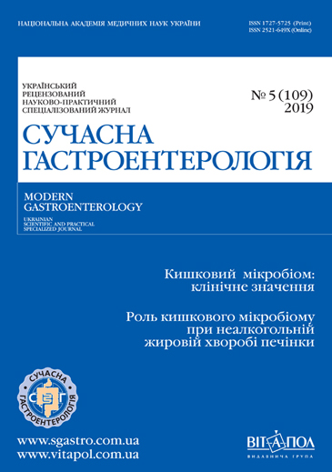
Неалкогольна жирова хвороба печінки в контексті кишкового мікробіому
DOI:
https://doi.org/10.30978/MG-2019-5-97Ключові слова:
неалкогольна жирова хвороба печінки, мікробіом кишечника, інсулінорезистентність, лікуванняАнотація
Наведено сучасні дані щодо епідеміологічної ситуації з неалкогольною жировою хворобою печінки (НАЖХП) в світі, її поширеності серед населення європейських країн, гендерні особливості. Висвітлено роль мікробіому кишечника в патогенетичних механізмах розвитку НАЖХП на стадії стеатозу, а також у прогресуванні захворювання. Показано вплив основних патогенетичних чинників (коротколанцюгових жирних кислот, жовчних кислот, ліпополісахаридів, зміни проникності кишкової стінки, співвідношення основних кластерів бактерій) на формування захворювання у здорових осіб та розвиток НАЖХП. Установлено взаємозв’язок інсулінорезистентності із зазначеними чинниками та їхній вплив на її значення у пацієнтів з метаболічним синдромом. Показано анатомічний і функціональний зв’язок кишечника та печінки в рамках кишкової мікробіоти. Представлено експериментальні дані, котрі доводять вплив кишкової мікробіоти на формування і прогресування неалкогольного стеатозу. Висвітлено вплив метаболітів кишкової мікробіоти (етанолу, жовчних кислот, триметиламін-N-оксиду, ароматичних амінокислот) на формування стеатозу печінки. Представлено експериментальні дані, котрі дали змогу пояснити вплив кишкової мікробіоти на вияви метаболічного синдрому і зокрема на НАЖХП, а також дані клінічних і преклінічних досліджень, присвячених вивченню впливу корекції мікробіому на провідні патогенетичні механізми НАЖХП. Розглянуто вплив окремих філотипів бактерій на перебіг НАЖХП. Висвітлено основні терапевтичні стратегії в лікуванні захворювання, котрі ґрунтуються на принципах доказової медицини, а також тенденції в антибіотикотерапії, терапії пробіотиками та фекальній трансплантації в контексті терапії НАЖХП та їхню потенційну роль.
Посилання
Boursier J, Mueller O, Barret M et al. The severity of nonalcoholic fatty liver disease is associated with gut dysbiosisand shift in the metabolic function of the gut microbiota. Hepatology. 2016;63:764-775.https://doi.org/10.1002/hep.28356
Bäckhed F, Ding H, Wang T et al. The gut microbiota as an environmental factor that regulates fat storage. Proc Natl Acad Sci USA. 2004;101:15718-15723.https://doi.org/10.1073/pnas.0407076101
Bajzer M, Seeley RJ. Physiology: obesity and gut flora. Nature. 2006;444:1009-1010.https://doi.org/10.1038/4441009a
Cohen NA. Novel indications for fecal microbial transplantation: update and review of the literature. Dig Dis Sci. 2017;62:1131-1145.https://doi.org/10.1007/s10620-017-4535-9
Cani P, Osto M, Geurts L. Involvement of gut microbiota in the development of low-grade inflammation and type 2 diabetes associated with obesity. Gut Microbes. 2012;3:279-288.https://doi.org/10.4161/gmic.19625
Compare D, Coccoli P, Rocco A et al. Gut—liver axis: the impact of gut microbiota on non alcoholic fatty liver disease. Nutr Metab Cardiovasc Dis. 2012;22:471-476.https://doi.org/10.1016/j.numecd.2012.02.007
Chalasani N, Younossi Z, Lavine J et al. The diagnosis and management of non-alcoholic fatty liver disease: practice Guideline by the American Association for the Study of Liver Diseases. Hepatology. 2012;55(6):2005-2023.https://doi.org/10.1053/j.gastro.2012.04.001
Del Chierico F, Nobili V, Vernocchi P et al. Gut microbiota profiling of pediatric nonalcoholic fatty liver disease and obese patients unveiled by an integrated meta-omics-based approach. Hepatology. 2017;65:451-464.https://doi.org/10.1002/hep.28572
Da Silva HE, Teterina A, Comelli EM et al. Nonalcoholic fatty liver disease is associated with dysbiosis independent of body mass index and insulin resistance. Sci Rep. 2018;8:1466.https://doi.org/10.1038/s41598-018-19753-9
Dao MC, Everard A, Wisnewsky A et al. Akkermansia muciniphila and improved metabolic health during a dietary intervention in obesity: relationship with gut microbiome richness and ecology. Gut. 2016;65:426-436.https://doi.org/10.1136/gutjnl-2014-308778
Di Luccia B, Crescenzo R, Mazzoli A et al. Rescue of fructose-induced metabolic syndrome by antibiotics or faecal transplantation in a rat model of obesity. PLoSONE. 2016;10:13-17.https://doi.org/10.1371/journal.pone.0134893
Fasano A, Physiological, pathological, and therapeutic implications of zonulin-mediated intestinal barrier modulation: living life on the edge of the wall. Am J Pathol. 2008;173:1243-1252.https://doi.org/10.2353/ajpath.2008.080192
Fracanzani AL, Valenti L, Bugianesi E et al. Risk of severe liver disease in NAFLD with normal aminotransferase levels: a role for insulin resistance and diabetes. Hepatology. 2008;48:792-798.https://doi.org/10.1002/hep.22429
Ferrere G, Wrzosek L, Cailleux F et al. Fecal microbiota manipulation prevents dysbiosis and alcohol-induced liver injury in mice. J Hepatol. 2016;66:806-815.https://doi.org/10.1016/j.jhep.2016.11.008
Gangarapu V, Ince AT, Baysal B. Efficacy of rifaximin on circulating endotoxins and cytokines in patients with nonalcoholic fatty liver disease. Eur J Gastroenterol Hepatol. 2015;27:840-845.https://doi.org/10.1016/s0016-5085(15)30129-3
Harte AL, da Silva NF, Creely SJ. Elevated endotoxin levels in non-alcoholic fatty liver disease. Journal of Inflammation (Lond). 2015;7:15.https://doi.org/10.1186/1476-9255-7-15
Hasan I. Perlemakan Hati Non Alkoholik. // Interna Publishingю–2009:695-701.https://doi.org/10.24293/ijcpml.v16i1.1027
Jandhyala SM, Talukdar R, Subramanyam C. Role of the normal gut microbiota. World J Gastroenterol. 2015;21:8787-8803.https://doi.org/10.3748/wjg.v21.i29.8787
Karagiannides I, Pothoulakis C. Obesity, innate immunity and gut inflammation. Curr Opin Gastroenterol. 2007;23:661-666.https://doi.org/10.1097/mog.0b013e3282c8c8d3
Kim B, Park K.-Y., Ji Y. Protective effects of Lactobacillus rhamnosus GG against dyslipidemia in highdiet-induced obese mice. Biochem Biophys Res Commun. 2016;473:530-536.https://doi.org/10.1016/j.bbrc.2016.03.107
Lofmark S, Edlund C, Jansson JK. Long-term ecological impacts of antibiotic administration on the human intestinal microbiota. ISME J. 2007;1:56-66.https://doi.org/10.1038/ismej.2012.91
Loomba R, Seguritan V, Li W et al. Gut microbiome-based metagenomic signature for non-invasive detection of advanced fibrosis in human nonalcoholic fatty liver disease. Cell Metab. 2017;25:1054-1062. https://doi.org/10.1016/j.cmet.2019.08.002
Le Roy T, Llopis M, Lepage P et al. Intestinal microbiota determines development of non-alcoholic fatty liver disease in mice. Gut. 2013;62:1787-1794.https://doi.org/10.1136/gutjnl-2012-303816
Li Z, Yang S, Lin H et al. Probiotics andantibodies to TNF inhibit inflammatory activity and improve nonalcoholic fatty liver disease. Hepatology. 2003;37:343-350.https://doi.org/10.1053/jhep.2003.50048
Ley RE. Obesity and the human microbiome. Curr Opin Gastroenterol. 2010;26:5-11.https://doi.org/10.1097/mog.0b013e328333d751
Lau E, Carvalho D, Freitas P. Gut microbiota: Association with NAFLD and metabolic disturbances. Biomed Res Int. 2015;97:95-99.https://doi.org/10.1155/2015/979515
Loomba R, Sanyal AJ. The global NAFLD epidemic. Nat Rev Gastroenterol Hepatol. 2013;Vol 10 (11):686-690.https://doi.org/10.1038/nrgastro.2013.171
Menni C, Fauman E, Erte I et al. Biomarkers for type 2 diabetes and impaired fasting glucose using a nontargeted metabolomics approach. Diabetes. 2013;62:4270-4276.https://doi.org/10.2337/db13-0570
Miele L, Valenza V, La Torre G. Increased intestinal permeability and tight junction alterations in nonalcoholic fatty liver disease. Hepatology. 2013;49:1877-1887.https://doi.org/10.1002/hep.22848
Mehta RS, Abu-Ali GS, Drew DA et al. Stability of the human faecalmicrobiome in a cohort of adult men. Nat Microbiol. 2018;3:347-355.https://doi.org/10.1038/s41564-017-0096-0
Machado MV, Cortez-Pinto H. Gut microbiota and nonalcoholic fatty liver disease. Ann Hepatol. 2012;11:440-449.https://doi.org/10.1016/s1665-2681(19)31457-7
Marchesini G, Mazzotti A. NAFLD incidence and remission: only a matter of weight gain and weight loss?. J Hepatol. 2015;62:15-17.https://doi.org/10.1016/j.jhep.2014.10.023
Okubo H, Sakoda H, Kushiyama A et al. Lactobacillus casei strain Shirota protects against nonalcoholic development in a rodent model. AJP Gastrointest Liver Physiol. 2013;305:911-918.https://doi.org/10.1152/ajpgi.00225.2013
Puri P, Daita K, Joyce A et al. The presence and severity of nonalcoholic steatohepatitis is associated with specific changes in circulating bile acids. Hepatology. 2018;67:534-548.https://doi.org/10.5604/01.3001.0011.7378
Rau M et al. Fecal SCFAs and SCFA-producing bacteria in gut microbiome of human NAFLD as a putative link to systemic T-cell activation and advanced disease. United European Gastroenterology Journal. 2018;6 (10):1496-1507.https://doi.org/10.1177/2050640618804444
Singh R, Sripada L. Side effects of antibiotics during bacterial infection: Mitochondria, the main target in host cell. Mitochondrion. 2014;16:50-54.https://doi.org/10.1016/j.mito.2013.10.005
Sabaté JM, Jouët P, Harnois F. High prevalence of small intestinal bacterial overgrowth in patients with morbid obesity: a contributor to severe hepatic steatosis. Obesity Surgery. 2007;18:371-377.https://doi.org/10.1007/s11695-007-9398-2
Sze MA, Schloss PD. Looking for a signal in the noise: Revisiting obesity and themicrobiome. MBio. 2016;7:1016-1018. https://doi.org/10.1128/mbio.01018-16
Sebastião MB. Duartea B, Claudia P. Microbiota and nonalcoholic fatty liver disease/nonalcoholic steatohepatitis (NAFLD/NASH). Annals of Hepatology. 2019;18:416-421.doi.org/10.1016/j.aohep.2019.04.006
Santacruz A, Collado MC, García-Valdés L et al. Gut microbiota composition is associated with body weight, weight gain and biochemical parameters in pregnant women. Br J Nutr. 2010;104:83-92.https://doi.org/10.1017/s0007114510000176
Vrieze A, Van Nood E, Holleman F et al. Transfer of intestinal microbiota from leandonors increases insulin sensitivity in individuals with metabolic syndrome. Gastroenterology. 2012;143:913-916.https://doi.org/10.1053/j.gastro.2012.06.031
Wu W. Small intestinal bacteria overgrowth decreases small intestinal motility in the NASH rats. World J Gastroenterol. 2008;14:313.https://doi.org/10.3748/wjg.14.313
Wong VW, Vergniol J, Wong GL et al. Diagnosis of fibrosis and cirrhosis using liver stiffness measurement in nonalcoholic fatty liver disease. Hepatology. 2010;51(2):454-462.
Winda P. Microbiota profiles in nonalcoholic fatty liver disease and its possible impact on disease progression evaluated with transient elastography: Lesson Learnt from 60 Cases. Сase Rep Gastroenterol. 2019;125:133.https://doi.org/10.1159/000498946
Wang J, Tang H, Zhang et al. Modulation of gut microbiota during probiotic-mediated attenuation of metabolic syndrome in high fat diet-fed mice. ISME. 2015;9:1-15.https://doi.org/10.1038/ismej.2014.99
Xu J, Gordon JI. Honor thysymbionts. Proc Natl Acad Sci USA. 2003;100:10452-10459.
Yatsunenko T, Rey FE, Manary MJ et al. Human gut microbiome viewed across age and geography. Nature. 2012;486:222-227.https://doi.org/10.3410/f.717247854.793478786
Zhu W, Gregory JC, Org E et al. Gut microbialmetabolite TMAO enhances platelet hyperreactivity and thrombosis risk. Cell. 2016;165:111-124.https://doi.org/10.1016/j.cell.2016.02.011
Zhao Y, Wu J. Gut microbiota composition modifies fecal metabolic profiles in mice. J Proteome Res. 2013;12:2987-2999.https://doi.org/10.1021/pr400263n
Zhu L, Baker RD, Zhu R. Gut microbiota produce alcohol and contributeto NAFLD. Gut. 2016;65:1-15.https://doi.org/10.1136/gutjnl-2016-311571


