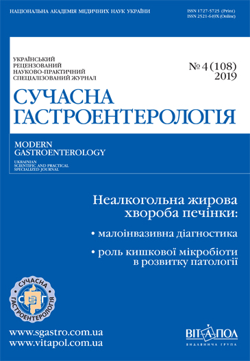
Автоімунний гастрит: проблеми діагностики та терапії
DOI:
https://doi.org/10.30978/MG-2019-4-59Ключові слова:
автоімунний гастрит, діагностика, лікування, перніціозна анемія, залізодефіцитна анеміяАнотація
Хронічний гастрит (ХГ) — одне з найпоширеніших захворювань внутрішніх органів, яким страждає до 20 % дорослої популяції. Сучасна парадигма ХГ розглядає захворювання як стартовий майданчик для формування раку шлунка, а також ерозивно-виразкових уражень, зокрема індукованих прийомом протизапальних препаратів. Широка поширеність симптомів шлункової диспепсії нерідко зумовлює гіпердіагностику ХГ, тоді як верифікація діагнозу має ґрунтуватися лише на клініко-ендоскопічних і патоморфологічних критеріях.
Останніми роками представлено великий масив наукових даних щодо ролі різних видів мікроорганізмів, зокрема бактерії H. pylori, дуоденогастрального рефлюксу, прийому медичних препаратів у розвитку і прогресуванні ХГ. Однак автоімунна форма цього захворювання є маловивченою. Загальновідомо, що автоімунний гастрит (АІГ) — це найважливіший чинник розвитку перніціозної анемії, а гіпохлоргідрія призводить до залізодефіцитної анемії. Роль АІГ у формуванні шлункової атрофії і метаплазії вивчено недостатньо, так само, як і гастроканцерогенний потенціал АІГ. На відміну від інших морфологічних форм ХГ діагностика АІГ потребує не лише визначення клініко-ендоскопічних і патогістологічних показників, а й аналізу серологічних даних. З позиції сучасних класифікацій ХГ (Кіотського консенсусу та системи OLGA/OLGIM) становить інтерес кореляція між серологічними і патогістологічними маркерами у пацієнтів з АІГ.
Лікування АІГ неспецифічне і принципово не відрізняється від терапії інших форм ХГ. Однак з огляду на патогенетичні особливості АІГ перспективним є розробка методів корекції імунопатологічних процесів, які характеризують це захворювання. Лікування АІГ спрямоване на ерадикацію інфекції, відновлення моторно-евакуаторної функції гастродуоденальної зони, нейтралізацію шкідливої дії жовчних кислот, однак стратегічним напрямом у терапії захворювання є гастропротекція. Використання препаратів на основі солей вісмуту сприяє відновленню структурного і функціонального стану слизової оболонки шлунка, регресу атрофії та кишкової метаплазії, гальмує прогресування АІГ.
Посилання
Zak MJu. Khronichnyy̆ ghastryt u khvorykh na osteoartroz: kliniko-proghnostychni, morfofunkcionaljni ta terapev- tychni aspekty: Avtoref. dys....d-ra med. nauk. Kharkiv,, 2017:41.
Zak MJu, Mosiy̆chuk LM. Khronichnyy̆ ghastryt i peredrak shlunka: Prakt. posibnyk. Dnipropetrovsjk, 2011:71.
Lyvzan MA, Mozghovoy̆ SY, Kononov AV. Ghastryt posle эradykacyy Helicobacter pylori — prostыe sledы yly serj’eznыe posledstvyja?. Suchasna ghastroenterol. 2011; 6 (62):73-76. Індексу DOI немає
Plotnykova EJu, Sukhykh AS. Preparatы vysmuta v praktyke vracha. Lechashhyy̆ vrach. 2016; 2.
Stuklov NY, Semenova EN. Zhelezodefycytnaja anemyja. Sovremennaja taktyka dyaghnostyky y lechenyja, kryteryy эffektyvnosty terapyy. Klyn. med. 2013;91,12; 12:61-67.
Kharchenko NV, Muzyka SV, Zak MJu. Peredrakovi zminy slyzovoï obolonky shlunka: mizhdyscyplinarnyy̆ poghljad na problem. Suchasna ghastroenterol. 2018; 4. DOI: 10.30978/MG-2018-3-96
Annibale B et al. Gastrointestinal causes of refractory iron deficiency anemia in patients without gastrointestinal symptoms. Am J Med. 2001;111(6):439-445. DOI:10.1016/s0002-9343 (01)00883-x
Annibale B, Lahner E, Fave GD. Diagnosis and management of pernicious anemia. Curr Gastroenterol Rep. 2011;13(6):518-524. DOI:10.1007/s11894-011-0225-5
Antico A et al. Clinical usefulness of the serological gastric biopsy for the diagnosis of chronic autoimmune gastritis. Clin Dev Immunol. 2012;2012:520970. DOI:10.1155/2012/520970
Bizzaro N, Tozzoli R, Shoenfeld Y. Are we at a stage to predict autoimmune rheumatic diseases?. Arthritis Rheum. 2007;56(6):1736-1744. DOI:10.1002/art.22708
Cabrera de Leon A et al. Factors associated with parietal cell autoantibodies in the general population. Immunol Lett. 2012;147 (1-2):63-66. DOI:10.1016/j.imlet.2012.06.004
Centanni M et al. Atrophic body gastritis in patients with autoimmune thyroid disease: an underdiagnosed association. Arch Intern Med. 1999;159 (15):1726-1730. DOI:10.1001/archinte.159.15.1726
Chanarin I. Pernicious anaemia as an autoimmune disease. Br J Haematol. 1972;23 (suppl.):101-107. DOI: 10.1111/j.1365-2141.1972.tb03510.× 14. Chiovato L et al. Disappearance of humoral thyroid autoimmunity after complete removal of thyroid antigens. Ann Intern Med. 2003;139 (5 Pt 1):346-351. DOI:10.7326/0003-4819-139-5_part_1-200309020-00010
Claeys D et al. The gastric H+/K+-ATPase is a major autoantigen in chronic Helicobacter pylori gastritis with body mucosa atrophy. Gastroenterology. 1998;115(2):340-347. DOI:10.1016/s0016-5085 (98)70200-8
De Block CE, De Leeuw IH, Van Gaal LF. Autoimmune gastritis in type 1 diabetes: a clinically oriented review. J Clin Endocrinol Metab. 2008;93(2):363-371. DOI:10.1210/jc.2007-2134
Dickey W et al. Gastric as well as duodenal biopsies may be useful in the investigation of iron deficiency anaemia. Scand J Gastroenterol. 1997;32(5):469-472. DOI:10.3109/00365529709025083
Ghosh T et al. Review article: methods of measuring gastric acid secretion. Aliment Pharmacol Ther. 2011;33(7):768-781. DOI: 10.1111/j.1365-2036.2010.04573.× 19. Gram K, Gram HC. Relations between achylia gastrica and simple and pernicious anemia. Arch Intern Med. 1924;34:658-668. DOI:10.1001/archinte.1924.00120050075004
Hershko C et al. Variable hematologic presentation of autoimmune gastritis: age-related progression from iron deficiency to cobalamin depletion. Blood. 2006;107(4):1673-1679. DOI:10.1182/blood-2005-09-3534
Hershko C, Patz J, Ronson A. The anemia of achylia gastrica revisited. Blood Cells Mol Dis. 2007;39(2):178-183. DOI:10.1016/j.bcmd.2007.03.006
Israeli E et al. Anti-Saccharomyces cerevisiae and antineutrophil cytoplasmic antibodies as predictors of inflammatory bowel disease. Gut. 2005;54(9):1232-1236. DOI: 10.1136/gut.2004.060228
Kaye PV et al. The clinical utility and diagnostic yield of routine gastric biopsies in the investigation of iron deficiency anemia: a case-control study. Am J Gastroenterol. 2008;103 (11):2883-2889. DOI:10.1111/j.1572-0241.2008.02121.x
Kulnigg-Dabsch S. Autoimmune gastritis. Wien Med Wochenschr. 2016;Bd. 166 (13):424-430. DOI: 10.1007/s10354-016-0515-5
Kulnigg-Dabsch S et al. Sa2034 Autoimmune Gastritis Is Common in Patients With Iron Deficiency - Non-Invasive Evaluation of Iron Deficiency Aside Guideline Recommendations. Gastroenterology. DOI: https:. doi.org/10.1016/S0016-5085 (15)31308-1
Lahner E et al. Reassessment of intrinsic factor and parietal cell autoantibodies in atrophic gastritis with respect to cobalamin deficiency. Am J Gastroenterol. 2009;104(8):2071-2079. DOI:10.1038/ajg.2009.231
McNicholl AG et al. Accuracy of GastroPanel for the diagnosis of atrophic gastritis. Eur J Gastroenterol Hepatol. 2014;26(9):941-948. [PMC free article] DOI:10.1097/MEG.0000000000000132
Neumann WL et al. Autoimmune atrophic gastritis — pathogenesis, pathology and management. Nat Rev Gastroenterol Hepatol. 2013;10(9):529-541. DOI:10.1038/nrgastro.2013.101
Okano A, Takakuwa H, Matsubayashi Y. Parietal-cell hyperplasia mimicking sporadic fundic gland polyps in the atrophic mucosa of autoimmune gastritis. Gastrointest Endosc. 2007;66(2):394-395. DOI:10.1016/j.gie.2007.01.022
Park JY, Lam-Himlin D, Vemulapalli R. Review of autoimmune metaplastic atrophic gastritis. Gastrointest Endosc. 2013;77(2):284-292. DOI: https:. doi.org/10.1016/j.gie.2012.09.033
Perros P et al. Prevalence of pernicious anaemia in patients with type 1 diabetes mellitus and autoimmune thyroid disease. Diabet Med. 2000;17 (10):749-751. DOI:10.1046/j.1464-5491.2000.00373.× 32. Pittman ME et al. Autoimmune metaplastic atrophic gastritis: recognizing precursor lesions for appropriate patient evaluation. Am J Surg Pathol. 2015;39 (12):1611-1620. DOI:10.1097/PAS.0000000000000481
Stolte M et al. Active autoimmune gastritis without total atrophy of the glands. Z Gastroenterol. 1992;30 (10):729-735.
Sugano K, Tack J, Kuipers EJ et al. Kyoto global consensus report on Helicobacter pylori gastritis. Gut. 2015;64(9):1353-1367. DOI:10.1136/gutjnl-2015-309252
Toh BH. Diagnosis and classification of autoimmune gastritis. Autoimmun Rev. 2014;13 (4-5):459-462. DOI: 10.1016/j.autrev.2014.01.048
Toh BH et al. Cutting edge issues in autoimmune gastritis. Clin Rev Allergy Immunol. 2012;42(3):269-278. DOI:10.1007/s12016-010-8218-y
Toh BH et al. Parietal cell antibody identified by ELISA is superior to immunofluorescence, rises with age and is associated with intrinsic factor antibody. Autoimmunity. 2012;45(7):527-532. DOI:10.3109/08916934.2012.702813
Toh BH, Sentry JW, Alderuccio F. The causative H+/K+-ATPase antigen in the pathogenesis of autoimmune gastritis. Immunol Today. 2000;21(7):348-354. DOI: 10.1016/S0167-5699 (00)01653-4
Tozzoli R et al. Autoantibodies to parietal cells as predictors of atrophic body gastritis: a five-year prospective study in patients with autoimmune thyroid diseases. Autoimmun Rev. 2010;10(2):80-83. DOI:10.1016/j.autrev.2010.08.006
Tozzoli R. The diagnostic role of autoantibodies in the prediction of organ-specific autoimmune diseases. Clin Chem. Lab. Med. 2008;46(5):577-587. DOI: 10.1515/CCLM.2008.138
Tursi A et al. Noninvasive prediction of chronic atrophic gastritis in autoimmune thyroid disease in primary care. Scand J Gastroenterol. 2014;49 (11):1394-1396. DOI: 10.3109/00365521.2014.958097
Uibo R et al. The relationship of parietal cell, gastrin cell, and thyroid autoantibodies to the state of the gastric mucosa in a population sample. Scand J Gastroenterol. 1984;19(8):1075-1080.
Van Herwaarden MA, Samsom M, Smout AJ. 24-h recording of intragastric pH: technical aspects and clinical relevance. Scand J Gastroenterol. 1999;230:9-16. DOI:10.1080/003655299750025219
Vannella L et al. Systematic review: gastric cancer incidence in pernicious anaemia. Aliment Pharmacol Ther. 2013;37(4):375-382. DOI:10.1111/apt.12177
Zhang Y et al. Gastric parietal cell antibodies, Helicobacter pylori infection, and chronic atrophic gastritis: evidence from a large population-based study in Germany. Cancer Epidemiol Biomarkers Prev. 2013;22(5):821-826. DOI:10.1158/1055-9965.EPI-12-1343


