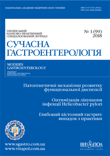
Функціональна диспепсія та імунна дисфункція: сучасні уявлення про патогенетичні механізми розвитку
Ключові слова:
функціональна диспепсія, імунна дисфункція, патогенез, дуоденальна еозинофілія, клітинний імунітет, гуморальний імунітетАнотація
Наведено сучасні погляди щодо імунологічних чинників розвитку функціональної диспепсії, дані про її мікроморфологічне підґрунтя. Імунну дисфункцію розглянуто як важливий чинник патогенезу функціональної диспепсії на різних рівнях — місцевому (у вигляді дуоденальної еозинофільної інфільтрації із дегрануляцією еозинофілів та мастоцитозу) та загальному (дисбаланс клітинного і гуморального імунітету з переважанням прозапальних цитокінів). Доведено перспективність подальшого вивчення ролі імунної регуляції в патогенезі функціональної диспепсії.Посилання
Andreev, D. N., et al. «Evolyutsiya predstavleniy o funktsionalnyih zabolevaniyah zheludochno-kishechnogo trakta v svete Rimskih kriteriev IV peresmotra (2016 g.).” Rossiyskiy zhurnal gastroenterologii, gepatologii, koloproktologii (Russian). 2017; 27 (1):4-11.
Babaeva, A. R., O. N. Rodionova, R. V. Vidiker. «Tsitokinovaya regulyatsiya funktsionalnyih zabolevaniy zheludochno-kishechnogo trakta.” Vestnik novyih meditsinskih tehnologiy (Russian). 2011;18 (1):163-164.
Koval, V. Yu., spivavt. «Klasyfikatsiia ta unifikovani klinichni protokoly pervynnoi, vtorynnoi (spetsializovanoi) medychnoi dopomohy pry zakhvoriuvanniakh stravokhodu ta shlunka.” (Ukrainian), 2015:63.
Martyinchuk AA, Gubskaya E.Yu. Sovremennyie perspektivyi diagnostiki i lecheniya pischevoy neperenosimosti. Likarska sprava. (Russian). 2014;11:64-68.
Palii, I. H. «Funktsionalna dyspepsiia: suchasni uiavlennia pro mekhanizmy vynyknennia y taktyku vedennia patsiientiv.” Praktykuiuchyi likar (Ukrainian). 2013;3:25-30.
Plotnikova, E. Yu., O. A. Krasnov. «Sochetanie funktsionalnoy dispepsii s gastroezofagealnoy reflyuksnoy boleznyu i hronicheskim gastritom.” Klinicheskie perspektivyi gastroenterologii, gepatologii (Russian). 2014;3:21-28.
Svintsitskyi, A. S., spivavt. «Morfolohichni zminy slyzovoi obolonky shlunka pry riznykh variantakh funktsionalnoi dyspepsii.” (Ukrainian) Morfolohiia. 2014;2:50-55.
Aro Pertti et al. Functional dyspepsia impairs quality of life in the adult population. Aliment Pharmacol Ther. 2011;N 33 (11):1215-1224.
Chen Ji, Yangde Zhang, Zhansheng Deng Imbalanced shift of cytokine expression between T helper 1 and T helper 2 (Th1/Th2) in intestinal mucosa of patients with post-infectious irritable bowel syndrome. BMC Gastroenterol. 2012;N 12 (1):91.
Cheung CK.Y. et al. Up-regulation of transient receptor potential vanilloid (TRPV) and down-regulation of brain-derived neurotrophic factor (BDNF) expression in patients with functional dyspepsia (FD). Neurogastroenterol Motil. 2017;N 30. DOI: 10.1111/nmo.13176.
Dizdar V et al. Relative importance of abnormalities of CCK and 5-HT (serotonin) in Giardia-induced post-infectious irritable bowel syndrome and functional dyspepsia. Aliment Pharmacol Ther. 2010;N 31 (8):883-891.
Dömötör András. et al. Immunohistochemical distribution of vanilloid receptor, calcitonin-gene related peptide and substance P in gastrointestinal mucosa of patients with different gastrointestinal disorders. Inflammopharmacol. 2005;N 13 (1):161-177.
Drossman DA. Functional gastrointestinal disorders: history, pathophysiology, clinical features, and Rome IV. Gastroenterol. 2016;N 150 (6):1262-1279.
Drossman DA, Hasler WL. Rome IV — functional GI disorders: disorders of gut-brain interaction. Gastroenterol. 2016;N 150 (6):1257-1261.
Du Lijun. et al. Corrigendum: increased duodenal eosinophil degranulation in patients with functional dyspepsia: a prospective study. Sci Rep. 2017;N 7.
Du Lijun. et al. Impact of gluten consumption in patients with functional dyspepsia: a case-control study. J Gastroenterol Hepatol. 2017.
Du Lijun. et al. Increased duodenal eosinophil degranulation in patients with functional dyspepsia: a prospective study. Sci Rep. 2016;N 6. doi: 10.1038/srep34305 (2016).
Farré R, Vicario M. Abnormal barrier function in gastrointestinal disorders. Gastrointest Pharmacol. 2017;N 1:193-217.
Ford AC et al. Global prevalence of, and risk factors for, uninvestigated dyspepsia: a meta-analysis. Gut. 2015;N 64:1049-1057.
Friesen CA et al. Eosinophils and mast cells as therapeutic targets in pediatric functional dyspepsia. World J Gastrointest. Pharmacol Ther. 2013;N 4 (4):86.
Futagami S, Itoh T, Sakamoto C. Systematic review with meta-analysis: post-infectious functional dyspepsia. Aliment Pharmacol Ther. 2015;N 41 (2):177-188.
Futagami S et al. Migration of eosinophils and CCR2-/CD68-double positive cells into the duodenal mucosa of patients with postinfectious functional dyspepsia. Am J Gastroenterol. 2010;N 105 (8):1835.
Kindt S et al. Intestinal immune activation in presumed post-infectious functional dyspepsia. Neurogastroenterol Motil. 2009;N 21 (8):832.
Kinoshita Y, Tsutomu C. and FUTURE Study Group Characteristics of Japanese patients with chronic gastritis and comparison with functional dyspepsia defined by ROME III criteria: based on the large-scale survey, FUTURE study. Internal Medicine. 2011;N 50 (20):2269-2276.
Kita H. Eosinophils: multifaceted biological properties and roles in health and diseas. Immunological Reviews. 2011;N 242 (1):161-177.
Koloski NA, Talley NJ, Boyce PM. The impact of functional gastrointestinal disorders on quality of life. Am J Gastroenterol. 2000;N 95 (1):67.
Lee Eun Hye, Hye Ran Yang, Hye Seung Lee Analysis of gastric and duodenal eosinophils in children with abdominal pain related functional gastrointestinal disorders according to Rome III criteria. J Neurogastroenterol Motil. 2016;N 22 (3):459.
Li Xiaobo. et al. The study on the role of inflammatory cells and mediators in post-infectious functional dyspepsia. Scand J Gastroenterol. 2010;N 45 (5):573-581.
Liebregts T et al. Small bowel homing T cells are associated with symptoms and delayed gastric emptying in functional dyspepsia. Am J Gastroenterol. 2011;N 106 (6):1089.
Manappallil RG, Thomas A. Clinical and endoscopic evaluation of dyspeptic patients attending a tertiary care hospital in South India: A prospective study. Asian Journal of Medical Sciences. 2017;N 8 (1):58-63.
Mosmann TR et al. Two types of murine helper T cell clone. I. Definition according to profiles of lymphokine activities and secreted proteins. J Immunol. 1986;N 136 (7):2348-2357.
Powell N, Walker MM, Talley NJ. The mucosal immune system: master regulator of bidirectional gut-brain communications. Nature Reviews Gastroenterol & Hepatol. 2017;N 14 (3):143-159.
Rodiño-Janeiro BK et al. Role of corticotropin-releasing factor in gastrointestinal permeability. J Neurogastroenterol Motility. 2015;N 21 (1):33.
Schmulson MJ, Drossman DA. What is new in Rome IV?. J Neurogastroenterol Motility. 2017;N 23 (2):151.
Schurman JV et al. Symptoms and subtypes in pediatric functional dyspepsia: relation to mucosal inflammation and psychological functioning. Journal of Pediatric Gastroenterology and Nutrition. 2010;N 51 (3):298-303.
Talley NJ. Functional dyspepsia: advances in diagnosis and therapy. Gut and liver. 2017;N 11 (3):349.
Talley NJ. Functional dyspepsia: new insights into pathogenesis and therapy. The Korean Journal of Internal Medicine. 2016;N 31 (3):444.
Talley NJ, Weaver AL, Zinsmeister AR. Impact of functional dyspepsia on quality of life. Digestive Diseases and Sciences. 1995;N 40 (3):584-589.
Talley NJ, Ford AC. Functional dyspepsia reply. New England Journal of Medicine. 2016;N 374 (9):896-896.
Talley NJ, Phillips SF. Non-ulcer dyspepsia: potential causes and pathophysiology. Ann Int Med. 1988;N 108 (6):865-879.
Talley NJ et al. Non-ulcer dyspepsia and duodenal eosinophilia: an adult endoscopic population-based case-control study. Clin Gastroenterol Hepatol. 2007;N 5 (10):1175-1183.
Vakil N. Functional dyspepsia — a disorder of duodenal permeability?. Aliment Pharmacol Ther. 2017;N 46 (1):70-71.
Vanheel H et al. Impaired duodenal mucosal integrity and low-grade inflammation in functional dyspepsia. Gut. 2014;N 63 (2):262-271.
Walker MM et al. Duodenal mastocytosis, eosinophilia and intraepithelial lymphocytosis as possible disease markers in the irritable bowel syndrome and functional dyspepsia. Aliment Pharmacol Ther. 2009;N 29 (7):765-773.
Walker MM et al. Implications of eosinophilia in the normal duodenal biopsy–an association with allergy and functional dyspepsia. Aliment Pharmacol Ther. 2010;N 31 (11):1229-1236.
Walker MM et al. The role of eosinophils and mast cells in intestinal functional disease. Current Gastroenterology Reports. 2011;N 13 (4):323-330.
Wallon C, Söderholm J. Corticotropin-releasing hormone and mast cells in the regulation of mucosal barrier function in the human colon. Ann NY Acad Sci. 2009;N 1165 (1):206-210.
Zheng Peng-Yuan. et al. Psychological stress induces eosinophils to produce corticotrophin releasing hormone in the intestine. Gut. 2009;N 58 (11):1473-1479.
Zheutlin LM et al. Stimulation of basophil and rat mast cell histamine release by eosinophil granule-derived cationic proteins. J Immunol. 1984;N 133 (4):2180-2185.
Zhu Jinfang, Hidehiro Yamane, Paul W. Differentiation of effector CD4 T cell populations. Ann Rev Immunol. 2009;N 28:445-489.


